COMPLETE DENTURE SUPPORT AND STABILITY
TABLE OF CONTENT
Define support, retention and stability
} To
state;
- Tissues
that provide primary and secondary support (in the maxilla and mandible)
- Factors
that influence the support of the dentures
- Factors
that affect retention
DENTURE SUPPORT
} It
is the resistance to vertical movement of the denture base towards the ridge at
right angles to the occlusal surfaces.
} The
tissues that provide this resistance are attached mucosa, the reflected mucosa
and the underlying bone.
} Support
is the principle that describes how well the underlying mucosa keeps the
denture from moving in the vertical plane towards the arch in question and thus
being excessively depressed and moving deeper into the arch.
} Initial
denture support is achieved by using impression procedures that provide optimal
extension and functional loading of the supporting tissues, which vary in their
resiliency.
} Long
term support is obtained by directing forces of occlusion towards those tissues
most resistant to remodeling and resorptive changes.
Primary support is provided in the;
Ø Maxilla;
Basal bone, horizontal portion of the palate.
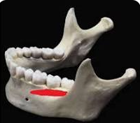
Ø Mandible; Pear shaped pad, buccal shelf.
Secondary support is provided by crest of the edentulous ridge.
} Factors
that influence support of the complete denture falls into 2 parts;
} Those
connected with supporting tissues.
} Those
connected with the denture and its base.
SUPPORTING TISSUE FACTORS
} They
are the foundation upon which the denture rests.
} They
are bone covered by mucosa and submucosa of varying thickness
NATURE OF SUPPORTING TISSUE
SOFT TISSUE:
} The
soft tissue should be firmly attached to the underlying cortical bone, it should
contain a resilient layer of submucosa and should be covered by keratinized
mucosa.
} The
soft tissue should not be flabby or hyperplastic.
HARD TISSUES:
} A
requirement of ideal support is the presence of tissues that are relatively
resistant to remodeling and resorptive changes.
} The
amount of bone loss after tooth loss is variable but greater in the mandible
than in the maxilla.
} The
horizontal plate of the hard palate lateral to the midline provides primary
support for complete denture.
} This
horizontal plate is resistant to resorption as it is covered by keratinized mucosa
and resilient submucosa.
} The
crest of the maxillary edentulous ridge is also important in support because
the soft tissue is often thick, keratinized and firmly bound to the periosteum.
} A
layer of dense fibrous connective tissue intervenes between the mucosa and the
bone and this connective tissue acts as a resilient liner for the mucosa.
} The
underlying cancellous bone is subject to resorptive changes so the alveolar
crest is considered as a secondary supporting area.
SUPPORTING AREA FOR THE LOWER DENTURE
} The
primary stress bearing area of the mandible must include the pear shaped pad
and the buccal shelf.
} The
pear shaped pad is the most distal extent of the keratinized masticatory
mucosa of the mandibular ridge.
} It
is different from the more distal retromolar pad which is alveolar mucosa,
overlying glandular and loose areolar connective tissue.
} The
retromolar pad is not a favorable denture bearing area. The junction of the
pear shaped pad and the retromolar pad demarcates the distal border of a
properly extended lower dentures.
} Buccal Shelf
} This
is a primary support area for the lower denture.
} It
is a thick buttress of bone projecting
from the mandible.
} It
is usually covered by the mucosa with an intervening submucosa layer which contains connective
tissue and buccinator muscle fibers.
} The
mandible ridge crest serves as the secondary support area because of lack of
muscle attachment and the presence of cancellous bone usually result in
resorptive changes occurring more rapidly than in the areas of primary support.
} Denture and its base
} Increase area coverage;
} Effective
support is realized by extension of denture base to cover maximal surface area
without impinging on movable tissues.
} To
reduce the load per unit area, one must increase the total area of the denture
base so that the load is spread over a large area without impinging on the
mobile tissues.
Area of occlusal table;
} A
patient will exert more force to penetrate food if the occlusal surfaces of the posterior teeth are broad
i.e if the teeth are big, more force
will be needed.
} The
use of posterior teeth that are narrow buccolingually will reduce the load on
the supporting tissues during mastication and increase the comfort of the
patient.
STABILITY
} Stability
is the resistance to horizontal or rotational forces.
} It
ensures the physiologic comfort of the patient.
} Lack
of stability often makes ineffective factors involved in retention and support.
} A
denture that shifts easily in response to laterally applied forces can cause
disruption of border seal or prevent the denture base from correctly relating
to the supporting tissue.
} The
ridge anatomy, base adaptation, residual ridge relationship, occlusal harmony,
neuromuscular control, all this can be condensed into the following category;
• Relationship
of denture base to the underlying tissues.
• Relationship
of external surface and border to the surrounding ororfacial musculature.
• Relationship
of opposing occlusal surfaces.
Ridge anatomy(height and form)
} Optimal
extension of denture base to contact movable tissues enhances stability (and
support)
} Relationship
of the external surface of the denture and its periphery to the surrounding
orofacial musculature.
} The
base adaptation to the residual ridge and the relationship of the polished
surface affects stability.
} Action
of the musculature on the base results in lateral and vertical dislodging
forces.
Complete Denture Stability can be enhanced by the following;
} Allowing
action of certain muscles group without the interference by the base so as not
to cause dislodging.
} Some
muscle function enhance stability.
} The
external surface of the denture should be developed to harmonize with the
following musculature of the lips, tongue and cheeks.
} The
buccal and labial flanges of upper and lower dentures should be concave to
permit seating by the cheeks and lips muscles.
} The artificial teeth are placed within the neutral zone.
} This
is an area where buccal and lingual forces generated by the musculature of the
lips, tongue and cheeks are balanced.
} It
is called the zone of minimal conflict.
} Relationship of the opposing occlusal surface
} The
denture must be free of interferences within the functional range of movement.
} The
functional range of movement refers to the position through which the jaw moves
horizontally during normal speech, swallowing and mastication.
} During
function, the occlusal surfaces should not make contact prematurely in
localized area.
} Lack
of occlusal balance causes the dentures to tilt on their supporting tissues
disrupting the retentive seal.
} The
anterior and posterior teeth should be arranged as close as possible to the
position once occupied by natural teeth.
} The
occlusal plane should not be too high. An elevated mandibular plane prevents
the tongue from reaching the food table into the sulcus.
} This
raised plane is caused by faults in occlusal vertical dimension causing
instability of dentures.
} Occlusal
balance plays a role in denture stability.


Post a Comment